CELLULAR IMAGING
EXPERTISE

A DEDICATED PLATFORM …
The Cellular Imaging platform of the Technology Centre provides expertise to the regional academic and private scientific community. The service is essentially organised around photonic microscopy techniques: wide field, confocal and HCS microscopy…
The platform operates under specific internal regulations, which are integrated into the internal regulations of the Technology Cluster and validated by the CRCT Laboratory Council.
The Cellular Imaging platform is located in the L2 containment area of the Technology Centre, on the 1st floor of the CRCT’s A building.
OUR EXPERTISE…
The technologies at our disposal offer a coherent set of tools for the study of fixed samples (cells or tissue fragments), either fluorescent or stained, or on samples studied in a dynamic manner.
Our missions:
Advice and assistance to users for acquisitions
Training of users in order to become autonomous
Development of research projects in the form of collaboration
Processing, image analysis, formatting and communication of results
Management of equipment

AVAILABLE DEVICES
The microscopes are freely available to users who have undergone compulsory training by the platform staff.
For other users, technical assistance is available by appointment.

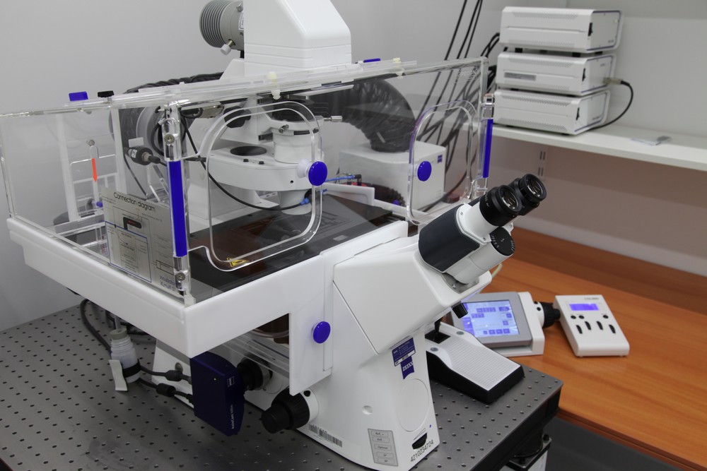
Confocal Zeiss LSM980 Airyscan 4Y
Microscope confocal inversé pour l’imagerie 3D sur des échantillons fixés (lame-lamelle) ou des échantillons vivants (support à fond lamelle de verre). Il permet l’amélioration de la résolution spatiale par le module AiryScan 2 et de la vitesse par l’option AiryScan Multiplex 4Y.

CONFOCAL LSM780 (Zeiss)
AxioObserver Z1 motorized inverted confocal microscope, it allows the realization of optical sections allowing resolved 3D reconstructions of fixed samples (slide-lamellar) or living samples (support with glass slide) and for F-techniques (FRAP, FLIM,)
Spinning-Disk / Vidéo-microscope
Microscope inversé avec un module spinning disk qui allie la rapidité du microscope champ large à la résolution d’un confocal.
Ce module permet des acquisitions d’images rapides en particulier sur des grands champs d’observation. Il permet des investigations sur cellules vivantes à des cadences rapides mais également des acquisitions sur de longues durées (jusqu’à 1 semaine d’observation).

OPERETTA CLS - High content Imaging system (Perkin elmar)
Automated inverted wide field fluorescence and confocal microscope (spinning disk). Ideal for measuring and analysing fixed or living cells in a controlled environment, with the ability to analyse multiple structural and functional cellular parameters in parallel.
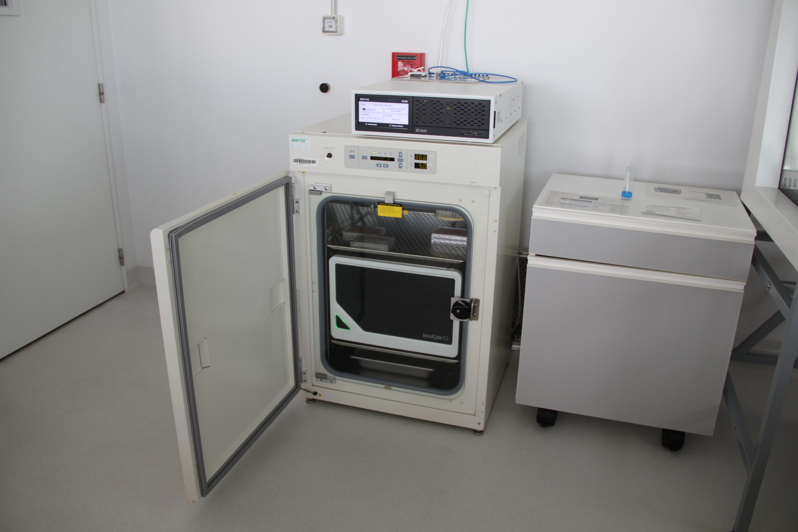
INCUCYTE S3
Automated imaging system that allows the monitoring of living cells in real time. The phenotype of the cells is associated live with various time-dependent quantitative data. Thanks to the analysis software, monitoring can be done 24 hours a day, over several days or even weeks, without any human intervention on the plates containing the cells under study. They remain confined under optimal conditions throughout the experiment.
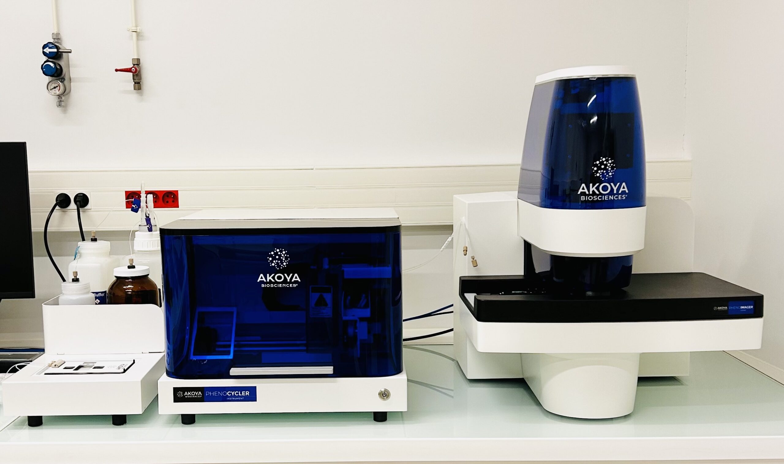
PHENOCYLCER-FUSION
The PhenoCycler-Fusion is an integrated platform that combines an automated cycling platform with a high-throughput imaging platform in an integrated end-to-end workflow.
INTERNAL REGULATION
All new users must have read the internal rules of the Cellular Imaging Platform and returned the information sheet.
MEMBERS OF THE PLATFORM
Laetitia Ligat
Ingénieur de laboratoire / laboratory engineer
Clément Cazorla
Ingénieur de laboratoire / laboratory engineer
Toulouse Cancer Research Center (Oncopole)
Toulouse - FR
Follow us on social network
Contact us
+33 5 82 74 15 75
Want to join
the CRCT team ?